Tooth Resorption in Cats: Causes, Symptoms & Treatment (Feline Resorptive Lesions)
If your cat has started tilting their head when eating, swallowing kibble without chewing, dropping food, or suddenly acting strange around the food bowl, it is often a sign that something in the mouth hurts.
One of the most common and painful reasons is tooth resorption – also called feline tooth resorption, feline resorptive lesions or FORLs. In this condition, a cat’s own body begins to break down and absorb parts of the tooth. Small “holes” appear, usually near the gumline, and they hurt a lot, even if your cat keeps eating.
Because cats are experts at hiding pain, tooth resorption is often missed until several teeth are affected. This guide explains what it is, how it is classified, what you can look for at home, how vets diagnose it, and which treatments actually help.
Quick Answer: What Is Tooth Resorption in Cats?
- Feline tooth resorption is a progressive dental disease where special cells (odontoclasts) break down a cat’s tooth structure.
- It typically affects the area where the tooth crown meets the root at the gumline, often starting below the gum.
- It is very common in pet cats and becomes more likely with age and with other dental disease.
- The American Veterinary Dental College (AVDC) recognizes three types of tooth resorption (Type 1, Type 2, Type 3) based on X-ray appearance, and five stages (1–5) based on severity.
- It is not the same as a cavity; it is not caused by bacteria but by the cat’s own cells.
- There is no medication, supplement or home remedy that can stop or reverse it. Treatment requires removing the painful tooth structure by extraction or crown amputation.
- There is currently no known way to prevent tooth resorption, but regular dental exams and dental X-rays allow early detection and timely treatment.
What Is Feline Tooth Resorption?
Tooth resorption happens when odontoclast cells, which normally help dissolve baby teeth so adult teeth can come in, become overactive in adult cats and target healthy permanent teeth instead.
Over time, these cells:
- Break down the outer surface of the tooth (enamel and cementum)
- Extend into the dentin and sometimes all the way into the pulp
- May cause the root to disappear and be replaced by bone-like tissue
From the outside, you might see:
- Small pits or holes at the gumline
- Pinkish areas where gum seems to grow into or onto the tooth
- Teeth that appear shortened, fractured or “missing” while roots remain below the gumline
Tooth resorption is not a cavity. Cavities come from bacterial acids dissolving enamel. Tooth resorption is the cat’s own body dissolving the tooth from inside or outside, often around the neck of the tooth.
Any tooth can be affected, but the lower premolars (cheek teeth) are especially common. Without treatment, this process leads to chronic pain.

How Common Is Tooth Resorption?
Tooth resorption is one of the most common dental diseases in cats. Studies in pet cats have found:
- Around 28.5–67% of cats have at least one resorptive lesion at some point in their life.
- The risk rises as cats get older.
- Cats with tartar, gingivitis and other dental disease are more likely to have tooth resorption.
- Some breeds have a noticeably higher risk, confirming a breed-specific predisposition.
A large case–control study published in 2024 showed strong associations between tooth resorption, age, dental disease and breed. Once a cat has had one lesion, it is much more likely to develop others in the future.
Because the disease usually starts below the gumline, many lesions are invisible without dental X-rays.
Types of Feline Tooth Resorption (T1, T2, T3)
The AVDC classifies tooth resorption into three main types based on how the roots and surrounding bone look on dental X-rays.
Type 1 (T1) – Inflammatory Resorption
- The crown of the tooth is damaged and resorbed.
- The root and periodontal ligament space (the thin dark line between root and bone) have a normal appearance on X-ray.
- Lesions often start at the neck of the tooth and are associated with inflammation, such as periodontal disease.
- The roots are still present and can remain painful if left behind.
Type 2 (T2) – Replacement Resorption
- The root appears to be disintegrating and is replaced with bone-like material.
- On X-ray, it becomes difficult to distinguish the root from the jawbone.
- The periodontal ligament space is narrowed or absent in affected areas.
- This is often a non-inflammatory process, and the root itself tends not to be painful in advanced T2 lesions.
Type 3 (T3) – Mixed Resorption
- A combination of Type 1 and Type 2 changes in the same tooth.
- Usually seen in multi-rooted teeth, where one root looks like Type 1 and another root looks like Type 2.
- Treatment decisions may differ for different roots of the same tooth, based on X-ray findings.
Summary Table – Types of Tooth Resorption
| Feature | Type 1 (T1) | Type 2 (T2) | Type 3 (T3) |
| X-ray appearance of root | Root normal and distinct from bone | Root blends with bone, hard to separate | One root T1, one root T2 in the same tooth |
| Periodontal ligament space | Normal width, visible | Narrowed or absent | Mixed: normal on some roots, absent on others |
| Association | Often with inflammation/periodontal disease | Often non-inflammatory, replacement process | Combination of both patterns |
| Typical treatment approach | Full extraction of crown and roots | Crown amputation when roots fully resorbing | Combination: extraction for T1 roots, T2 approach where appropriate |
Correctly identifying the type using dental X-rays is essential, because it guides how each tooth should be treated.
Stages of Tooth Resorption (1–5)
Tooth resorption is also staged based on how far the process has gone.
Stages of Feline Tooth Resorption
| Stage | What Happens |
| Stage 1 | Small defect on the outer surface of the tooth (enamel or cementum only). |
| Stage 2 | Lesion extends into dentin (the yellow, inner tissue) but not yet into the pulp. |
| Stage 3 | Lesion reaches the pulp cavity where nerves and blood vessels are located. |
| Stage 4 | Most of the tooth, including crown and root, is structurally destroyed. |
| Stage 5 | Tooth is almost completely resorbed; only small remnants remain on X-ray. |
Type (T1, T2, T3) and stage (1–5) together determine the best treatment for each tooth.
What Causes Tooth Resorption?
The underlying cause of feline tooth resorption is still unknown, and many cases are considered idiopathic. However, several factors are thought to contribute:
- Overactivation of odontoclasts that normally dissolve baby teeth
- Chronic inflammation in the mouth (especially in Type 1 lesions)
- Periodontal disease and gingivitis
- Possible diet and mineral imbalances, including vitamin D and calcium–phosphorus balance
- Breed and genetic predisposition
- Mechanical stress on teeth, misalignment, or frequent vomiting
Importantly:
- Tooth resorption has not been proven to be caused by bacteria, and it is not a classic cavity.
- There are no proven preventive diets, supplements, or oral products that specifically stop tooth resorption.
The strongest patterns are increased age, presence of other dental disease, and a higher long-term risk in cats that have already developed one lesion.
Symptoms: What You May Notice at Home
Cats are very good at hiding pain, so signs are often subtle or easy to misinterpret as “getting older” or “being picky.”
Common Symptoms of Tooth Resorption in Cats
| Category | Signs You May Notice |
| Eating changes | Dropping food, chewing on one side, swallowing kibble whole, preferring wet food, running away from the bowl after trying to eat |
| Pain behaviours | Jaw chattering (especially when the tooth is touched), drooling, pawing at the mouth, head shaking or tilting |
| Behaviour changes | Reduced grooming, dull or messy coat, irritability, hiding more, less interest in play and interaction |
| Visible mouth signs | Pink spots or small “holes” near the gumline, gum tissue growing over the tooth, fractured or missing tooth crowns |
Many cats show no obvious changes at home until lesions are advanced. Any combination of these signs is a reason to schedule a veterinary dental exam.
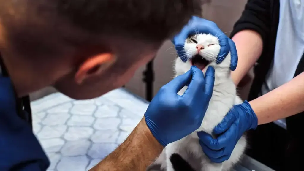
How Vets Diagnose Tooth Resorption
A complete diagnosis of tooth resorption cannot be made from a quick look in an awake cat’s mouth. Many lesions are below the gumline and invisible without imaging.
Diagnosis usually includes:
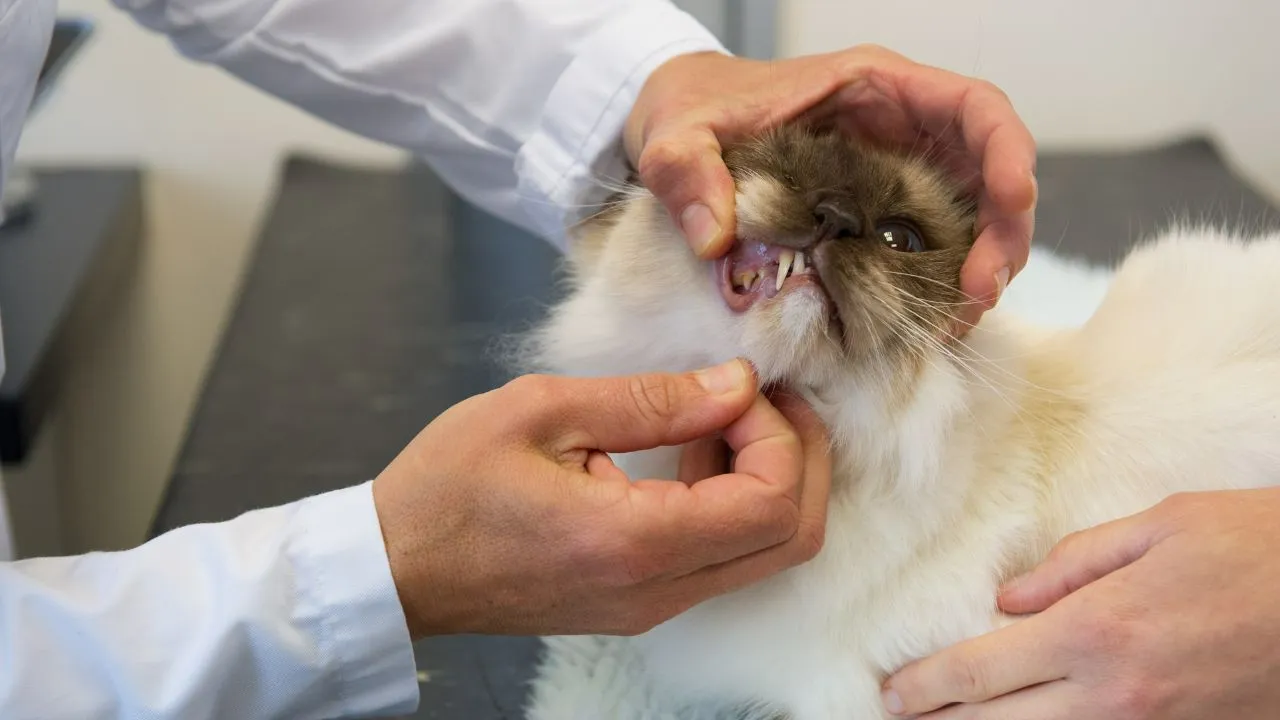
- Clinical oral exam
The vet examines the mouth, looks for inflammation, masses, ulcers and other oral diseases that can cause pain, such as gingivostomatitis or eosinophilic lesions. - Sedation or general anesthesia
To fully examine all teeth, probe around the gumline and avoid causing pain or fear, cats are examined under sedation or general anesthesia. - Dental X-rays (radiographs)
Full-mouth dental X-rays are essential to:- Identify lesions below the gumline
- Classify lesions as Type 1, Type 2 or Type 3
- Evaluate each root and the surrounding bone
- Plan appropriate treatment for each individual tooth
Often, additional lesions are discovered only once dental X-rays are done, especially when a cat is under anesthesia for a routine dental cleaning.
Treatment: How Tooth Resorption Is Managed
There are no non-surgical products, drops, gels or supplements that can stop or reverse tooth resorption. The aim of treatment is to remove the painful tooth structure and restore comfort.
Overview of Treatment by Type
| Lesion Type | Main Treatment | Notes |
| Type 1 (T1) | Full extraction of crown and roots | Roots remain intact and must be completely removed |
| Type 2 (T2) | Crown amputation when appropriate | Crown removed; resorbing roots left in bone if confirmed non-painful |
| Type 3 (T3) | Mixed approach | Some roots extracted as T1, others treated as T2 based on X-ray |
Surgical Extraction
- Most often used for Type 1 lesions and mixed cases.
- The entire tooth, including all roots, is carefully removed.
- Local nerve blocks are used to numb the area and reduce the amount of anesthesia needed.
- Dental X-rays confirm that no painful root fragments are left behind.
Crown Amputation (Coronal Amputation)
- Used selectively for Type 2 lesions where the root has fused with bone and is being replaced by bone-like tissue.
- The crown is removed at or below the gumline and the area is smoothed.
- Gum tissue is sutured over the site.
- The root remnants continue to resorb within the bone and are usually not painful.
Crown amputation should only be performed when high-quality dental X-rays confirm Type 2 resorption and when the procedure is done by a veterinarian trained in dental surgery.
Pain Management and Aftercare
Before, during and after treatment, pain control is essential:
- Local nerve blocks (for example with lidocaine or bupivacaine)
- Anti-inflammatory medications when appropriate
- Additional pain medications according to the vet’s judgment
After surgery, most cats eat soft food comfortably within a short time and then gradually return to their usual diet as healing progresses.
Palliative Care
In cats that cannot safely undergo anesthesia due to severe illness or advanced age, palliative care may be considered. This focuses on:
- Pain relief
- Softer diets
- Maintaining quality of life for as long as possible
Palliative care does not stop the disease but can temporarily improve comfort.
Life After Extractions: Can Cats Eat Without Teeth?
Most cats feel significantly better after painful teeth are removed. Many owners notice:
- More enthusiasm about food
- Improved grooming and a cleaner coat
- Increased affection and interaction
- Weight gain if the cat had been eating less
Cats can cope very well with missing teeth. Even cats with many extractions often learn to eat both soft food and, in some cases, dry food once the mouth has healed. Removing painful teeth is an act of kindness, not cruelty.
Can Tooth Resorption Be Prevented?
At this time, there is no proven way to prevent feline tooth resorption. However, you can improve overall dental health and catch problems earlier by:
- Scheduling regular veterinary dental check-ups
- Having professional dental cleanings done under anesthesia when recommended
- Allowing full-mouth dental X-rays when your vet advises them
- Brushing your cat’s teeth with cat-safe toothpaste if your cat will tolerate it
- Using veterinary-approved dental diets or chews to support general dental health
These measures help reduce plaque, gingivitis and periodontal disease, and make it more likely that tooth resorption is discovered earlier, before your cat has lived with severe pain for a long time.
FAQ: Tooth Resorption in Cats
Is tooth resorption the same as a cavity?
No. Cavities are caused by bacteria and acid damaging the tooth from the outside. Tooth resorption is caused by the cat’s own cells breaking down the tooth, often starting at or below the gumline.
Is tooth resorption painful?
Yes. Once the lesion reaches the dentin or pulp, it is very painful. Cats may still eat, but they adapt and hide discomfort.
Can tooth resorption heal on its own?
No. It is a progressive disease. Without proper treatment, it continues to advance and causes ongoing pain.
Are there drops, gels or supplements that cure tooth resorption?
No. There are no proven non-surgical products that stop or reverse tooth resorption. The only effective long-term treatment is removal of the affected tooth structure.
Will my cat need more teeth removed in the future?
Very likely. Once a cat has developed one lesion, the chances of developing others later are high. Regular dental exams and X-rays are important throughout life.
When should I call the vet?
Any signs of drooling, difficulty chewing, dropping food, jaw chattering, blood from the mouth, head tilting, pawing at the face or changes in eating habits and behaviour should be checked by a vet, preferably with a dental focus.